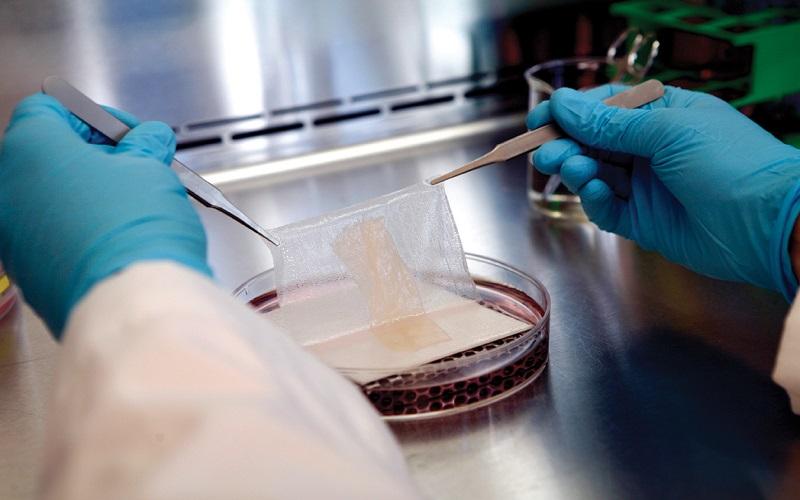
Frozen in Time is the story of how the answers to a few of life’s most difficult mysteries were found. Protein extraction from OCT embedded tissue samples is complicated by polymers that interfere with the MS signal (Supplementary Figure 1). This can be resolved with a simple cleaning step using diethyl ether/methanol.
Preparation
A key benefit of OCT Compound Tissue Freezing is its ability to preserve tissue structure in a block format before freezing, which is ideal for tissue microarray studies. This approach overcomes antigenic changes in proteins and mRNA degradation associated with paraffin-embedded specimens.
One disadvantage of OCT is the water soluble synthetic polymers it contains, which can cause ion suppression during MS analysis9. To mitigate this effect, Varna et al developed an optimized method for protein extraction from OCT embedded tissue samples. They removed the OCT from the tissue sample before thawing and performed methanol/chloroform/water extraction to remove residual OCT. The result was a higher protein yield compared to OCT samples without the removal step. The use of the OCT removal technique could significantly improve future proteomic analyses of frozen OCT-embedded tissue.
Freezing
Optimal cutting temperature (OCT) compound is an excellent embedding medium for frozen tissue specimens. Its clear solution is easy to use and provides superior sectioning at cryostat temperatures as low as -10degC. It also leaves no residue on slides, eliminating undesirable background staining that can occur with some other freezing compounds. It is available in a convenient 4 oz. spout plastic container.
Once the OCT is completely mixed and ready for use, add it to the cryomold in which the sample is being inserted. If the sample is larger than the mold, additional OCT may be added to cover the tissue. Once the OCT covers the tissue, the mold is placed in a liquid nitrogen Dewar and allowed to freeze. Once the OCT has fully frosted, transfer the frozen OCT block to a dry ice container and store in a -80 ultra-low freezer for biobanking.
Sectioning
Tissue snap frozen in OCT compound and sectioned on a cryostat yields sections with high quality, fine detail and excellent morphologic preservation. OCT Compound Tissue Freezing has good adhesion to slides and does not separate during the sectioning process. It is also compatible with chromogenic IHC reagents and does not dull microtome knives. Unlike some freezing compounds, OCT can be used multiple times without loss of quality. It is a water-soluble blend of glycols and resins that is clear, free of particles and does not leave residue on the slides during staining. It is easy to use, and is available in a convenient 4 oz. spout plastic bottle.
Once the tissue is surrounded by OCT compound and placed in the appropriate size cryomold, the sample should be gently agitated to ensure a flat surface is achieved for sectioning. OCT should be poured slowly over the sample to avoid bubble formation until all of the tissue is covered. After OCT is fully drained, the cryomold should be capped and placed on dry ice for quick freezing. After freezing, the OCT block should be transferred to a liquid nitrogen or mechanical -80 C freezer for long term storage.
Storage
The tissue pieces must be carefully placed in the OCT solution, making sure not to crowd the mold. Once the OCT is completely covering the tissue piece, it is ready to be frozen. It may take 0.5-1 minute for the OCT to harden. When it is done, the block of tissue should be tightly wrapped in aluminum foil and stored in a labeled freezer.
It is recommended to store the OCT Compound Tissue Freezing blocks upside down in order to prevent bubble formation during storage. This will help to avoid potential damage to the specimen during its storage.
Embedding of fresh, unfixed tissue samples in optimal cutting temperature (OCT) compound is an excellent procedure to use for snap freezing samples for later microtome sectioning. It is simple, fast, and allows for preservation of the primary morphology of the sample. In addition, it allows for the investigation of proteins in the resulting sections using mass spectrometry based proteomics. Thank visiting foxbopsot.com
Also Read: Rigid Boxes
